
Mastering Neuroimaging: A Beginner’s Guide for Medical Students
Discover the fundamentals of neuroimaging, from CT scans to MRIs, and learn how they’re used in diagnosing brain conditions.
This guide explains when to use CT vs MRI, what different scans reveal, and how to interpret basic findings. Perfect for clinical rotations or exam prep, it also includes key tips for understanding radiology reports.
Introduction:
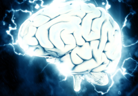
Neuroimaging is an essential skill for medical students, especially in the field of neurology and neurosurgery. This post will introduce you to the basics of neuroimaging and its importance in diagnosing neurological conditions.
Key Insights:
- MRI (Magnetic Resonance Imaging): Ideal for visualizing soft tissue, commonly used in brain and spinal cord imaging.
- CT (Computed Tomography): Quick method for detecting brain hemorrhages, fractures, and tumors.
- fMRI (Functional MRI): Assesses brain activity by measuring blood flow during task performance.
- PET (Positron Emission Tomography): Used to observe metabolic processes in the brain and detect abnormal tissue.
- Techniques in Practice: Understand how to interpret neuroimaging results in a clinical setting.
Conclusion:
As a medical student, gaining proficiency in neuroimaging will enhance your diagnostic capabilities. Learn to interpret these imaging techniques and their clinical applications to become a well-rounded healthcare professional.